• About
  • Subscribe
  • Advertise
  • Contact
Thursday, December 5, 2024
Newsletter
SUBSCRIBE
  • Latest News
  • All Sections
    • Ophthalmic insights
      • Policy & regulation
      • Company updates & acquisitions
      • Research
      • Clinical trials
      • Workforce
      • Product approvals
      • Conferences
      • Opinion
      • Indigenous eye health
      • Retail
    • Eye disease
      • Dry eye
      • Myopia
      • Cataract
      • Glaucoma
      • Macular disease – AMD
      • Diabetic eye disease
      • Inherited retinal disease
      • Corneal disease
      • Presbyopia
      • Eye infections
    • Ophthalmic Careers
      • New appointments
      • Industry profiles
      • Graduates
    • Ophthalmic organisations
      • Regulators
      • Optometry networks
      • Private ophthalmology clinics
      • Associations
      • Patient support bodies
      • Eye research institutions
      • Optometry schools
      • Optical Dispensing trainers
      • Medical schools
      • RANZCO
  • Features
    • Report
    • Soapbox
  • Ophthalmic education
    • CPD – Optometry
    • Optical Dispensing
    • Orthoptics Australia
    • Practice management
  • Products
    • Ophthalmic Treatments
      • Ophthalmic lenses
      • Lens treatments
      • Myopia interventions
      • Light-based therapy
      • Minimally invasive glaucoma surgery (MIGS)
      • Gene therapy
      • Laser treatments
      • Supplements
      • Eyewear & frames
      • Behavioural optometry/vision training
      • Contact lenses
      • Anti-VEGF
      • Intraocular lenses (IOLs)
      • Pharmaceuticals & consumables
    • Ophthalmic equipment & diagnostics
      • Biometry – axial length
      • Perimetry & visual fields
      • OCT
      • Phoropter
      • Autorefractor
      • Tonometry
      • Topography
      • Multimodal imaging
      • Retinal imaging
      • Anterior segment imaging
      • Software & data management
      • Microscopes
      • Slit lamps
      • Lens edging
      • Stands, chairs and tables
      • Ultrasound
      • Dry eye diagnostics
      • Low vision aids
  • Research
  • Classifieds
No Results
View All Results
Home Feature

Intraocular lymphomas … and sending specimens to the lab – Prof Sarah Coupland

by Staff Writer
March 29, 2023
in Feature, Soapbox
Reading Time: 4 mins read
A A
Intraocular lymphomas Prof Sarah Coupland

Figures of vitreoretinal lymphoma: the image shows a cytospin of a vitrectomy sample, stained with MGG (left) with atypical cells. On the right (in brown, using immunohistochemistry) is the CD20 stain highlighting the neoplastic B-cells.

Share on FacebookShare on Twitter


Intraocular lymphomas arise in different parts of the eye and show varied clinical manifestations, masquerading as many diseases; therefore, making the diagnosis is challenging.

Even when lymphoma is suspected, it can be difficult to confirm cytologically, because of the fragility of lymphoma cells. For these reasons, close cooperation between the ophthalmologist and pathologist is essential, particularly to optimise the information that can be obtained from these paucicellular samples.

Prof Sarah Coupland.

Such collaboration requires the ophthalmologist to have a good understanding of intraocular lymphoma pathology and basic knowledge of the laboratory investigations required.

In the first part of this Soapbox, we describe the clinical manifestations of intraocular lymphomas. In the second section, we make recommendations for sample transport and outline briefly the tests undertaken for their diagnosis.

Intraocular lymphoma types 

Essentially, intraocular lymphomas can be divided into: a) vitreoretinal lymphomas (VRL); b) primary choroidal lymphomas (PCL); c) secondary choroidal lymphomas. Rarer subtypes can occur as primary or secondary tumours in the iris; however, they will not be discussed here.

VRL are high-grade B-cell lymphomas that arise in the retina, with/without vitreous involvement. They typically are associated with central nervous system (CNS) disease, which can occur either concurrent or after the ocular disease.

In the new World Health Organization (WHO) Lymphoma classification, VRL has been recognised as a lymphoma-type that arises in an immune privileged site, alongside CNSL and testicular lymphoma. In general, VRL patients have a poor prognosis, particularly if diagnosis is delayed.

PCL are low-grade B-cell lymphomas, that respond very well to low-dose radiotherapy. They are very similar to conjunctival low-grade B-cell lymphomas. However, PCL can be extensive leading to retinal detachment; as well as infiltrate other parts of the uvea, mimicking a diffuse melanoma, or even spread into extraocular tissues.

Secondary choroidal lymphomas represent an intraocular manifestation of a systemic lymphoma. These typically occur in patients with advanced or relapsed Non-Hodgkin Lymphomas (NHL), with the subtypes varying considerably. The response to chemotherapy is dependent on the subtype of NHL.

Lab-based testing

Reaching the diagnosis of intraocular lymphoma requires good clinicopatholog- ical correlation. Communication between clinical and pathology lab teams is critical; particularly, it is important for the lab when the sample will be sent, avoiding late Friday afternoons, where possible.

Clinicians frequently-asked-questions concerns use of steroids prior to vitrectomy (best avoided) and the sample transport media of the vitrectomy or aspirate specimens.

We recommend a soft-fixative, such as CytoLyt or H.O.P.E. fixation, for both the undiluted and diluted samples. The cassette-washings should also be sent, as sometimes these contain the diagnostic cells.

The above-mentioned fixatives help maintain tumour cell integrity, and allow for same-day rapid cytospin preparations, and for most (if not all) laboratory tests (including molecular) to be undertaken.

Glutaraldehyde fixation should be avoided as it leads to poor quality DNA on extraction. Neutral buffered formalin for vitrectomy samples is not ideal as it requires wax-embedding, with the biopsied cells ‘settling’ at differing levels within the block. Numerous sections must then be undertaken to obtain a complete picture of the vitrectomy sample, often leaving insufficient material for essential molecular testing. However, it must be noted that should a chorioretinal biopsy be taken and is visible as a tissue piece, then this should be sent in buffered formalin.

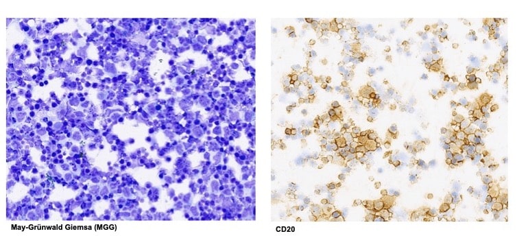
In addition to simple morphology stains, the main tests that are performed include immunohistochemistry (IHC), immunoglobulin chain rearrangement testing using polymerase chain reaction (IgH-PCR) and increasingly mutational gene analysis, e.g., MYD88.

The antibodies typically used in IHC include ones directed against: B-cells (CD79a, CD20 and PAX5), T-cells (CD3) and macrophages (CD68). Cells that are morphologically atypical and positive for a B-cell marker are highly suspicious of a B-cell lymphoma.

If these are within a vitrectomy sample and are medium-to-large in size, then this would speak for VRL. If these cells are smaller and the dominant population within a choroidal biopsy or aspiration, then this be suspicious of a primary choroidal lymphoma.

The molecular testing demonstrating either a monoclonal B-cell population and/or a MYD88 mutation (present in 70% of all VRL), steers diagnosis towards malignancy rather than inflammation. Integration of all findings is imperative to reach a definitive diagnosis.

NOTE: This article was co-authored by Dr Yamini Krishna, consultant histopathologist.

ABOUT THE AUTHOR
Name: Professor Sarah Coupland
Qualifications: MBBS, PhD, FRCPath
Primary place of work: Liverpool Clinical Laboratories and University of Liverpool
Position: Consultant Histopathologist and George Holt Chair of Pathology
Location: Liverpool, UK
Years in profession: 20 years

More reading

A proactive approach for glaucoma suspects – Dr Brian Ang

Port delivery system for neovascular AMD – A/Prof Anthony Kwan

Eye injuries and why sporting eyewear matters – Dr Gizem Ashraf

Related Posts

As a practice’s most valued asset, staff need training to ensure the business runs effectively. Image: YAKOBCHUK VIACHESLAV/Shutterstock.com.

Building a strong staff complement – Part 2

by Staff Writer
December 4, 2024

Having a structured approach to the employee lifecycle can set clear expectations and drive staff performance, writes Karen Crouch in...

A Google My Business account is the way practice owners can claim ownership of their business profile. Image: Vladimka production/Shutterstock.com.

Keeping on top of your business directory listings

by Staff Writer
December 3, 2024

Unknown to many practitioners, Google, Microsoft, and Apple have created business directory listings for them without their input or permission....

A low vision patient using an aid to check prices at the supermarket. Image: Vision Australia.

Low vision patients and the moments that matter

by Staff Writer
December 2, 2024

Simply spending a few minutes at the end of a consultation to discuss options with a low vision patient can...

Join our newsletter

Insight has been the leading industry publication in Australia for more than 40 years. This longevity is largely due to our ability to consistently deliver accurate and independent news relevant to all ophthalmic professionals and their supporting industry.

Subscribe to our newsletter

About Insight

  • About
  • Advertise
  • Subscribe
  • Contact
  • Terms & Conditions
  • Privacy Collection Notice
  • Privacy Policy

Popular Topics

  • Business
  • Feature
  • Research
  • Technology
  • Therapies
  • Classifieds

© 2024 All Rights Reserved. All content published on this site is the property of Prime Creative Media. Unauthorised reproduction is prohibited

No Results
View All Results
NEWSLETTER
SUBSCRIBE
  • Latest News
  • All Sections
    • Ophthalmic insights
      • Policy & regulation
      • Company updates & acquisitions
      • Research
      • Clinical trials
      • Workforce
      • Product approvals
      • Conferences
      • Opinion
      • Indigenous eye health
      • Retail
    • Eye disease
      • Dry eye
      • Myopia
      • Cataract
      • Glaucoma
      • Macular disease – AMD
      • Diabetic eye disease
      • Inherited retinal disease
      • Corneal disease
      • Presbyopia
      • Eye infections
    • Ophthalmic Careers
      • New appointments
      • Industry profiles
      • Graduates
    • Ophthalmic organisations
      • Regulators
      • Optometry networks
      • Private ophthalmology clinics
      • Associations
      • Patient support bodies
      • Eye research institutions
      • Optometry schools
      • Optical Dispensing trainers
      • Medical schools
      • RANZCO
  • Features
    • Report
    • Soapbox
  • Ophthalmic education
    • CPD – Optometry
    • Optical Dispensing
    • Orthoptics Australia
    • Practice management
  • Products
    • Ophthalmic Treatments
      • Ophthalmic lenses
      • Lens treatments
      • Myopia interventions
      • Light-based therapy
      • Minimally invasive glaucoma surgery (MIGS)
      • Gene therapy
      • Laser treatments
      • Supplements
      • Eyewear & frames
      • Behavioural optometry/vision training
      • Contact lenses
      • Anti-VEGF
      • Intraocular lenses (IOLs)
      • Pharmaceuticals & consumables
    • Ophthalmic equipment & diagnostics
      • Biometry – axial length
      • Perimetry & visual fields
      • OCT
      • Phoropter
      • Autorefractor
      • Tonometry
      • Topography
      • Multimodal imaging
      • Retinal imaging
      • Anterior segment imaging
      • Software & data management
      • Microscopes
      • Slit lamps
      • Lens edging
      • Stands, chairs and tables
      • Ultrasound
      • Dry eye diagnostics
      • Low vision aids
  • Research
  • Classifieds
  • About Us
  • Advertise with Insight
  • Subscribe
  • Contact Insight

© 2024 All Rights Reserved. All content published on this site is the property of Prime Creative Media. Unauthorised reproduction is prohibited